Indolent Ulcers in Dogs: Signs, Treatment, and Recovery
Indolent corneal ulcers are painful, non-healing eye wounds in dogs that require veterinary treatment to help the cornea heal.
An indolent corneal ulcer is a surface wound of the eye that won’t heal the way a typical scratch would. The surface layer of the cornea has trouble attaching to the tissue beneath it, which can leave the eye painful and slow to heal without veterinary treatment. Although indolent ulcers are superficial, they still require veterinary care to prevent complications that could threaten your dog’s comfort and vision.
What Is an Indolent Corneal Ulcer?
The cornea is the clear outer surface of the eye. It has several layers, including the epithelium, which forms its protective outer layer.
With an indolent corneal ulcer, the epithelium does not attach normally to the tissue underneath it. As a result, the ulcer can remain open even though there is not a deeper injury to the cornea. A loose edge of the corneal epithelium may be visible around the ulcer.
Indolent corneal ulcers are most often diagnosed in middle-aged to older dogs. They can occur in any breed, although some breeds, including Boxers and French Bulldogs, appear to be more commonly affected.
These ulcers are sometimes called recurrent corneal ulcers or recurrent corneal erosions. However, an ulcer does not have to occur more than once to be considered indolent.
Signs and Diagnosis
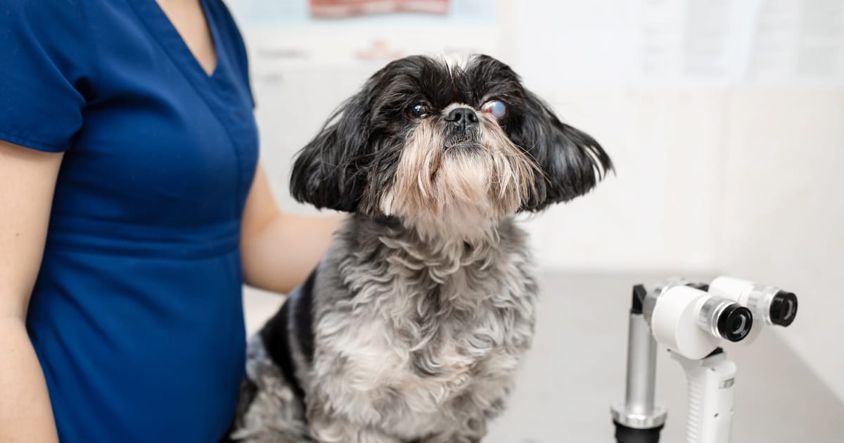
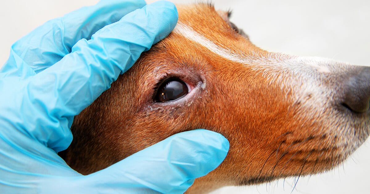
Corneal ulcers can be painful, and dogs may show several signs when an ulcer is present. Squinting or keeping the affected eye closed is common, along with excessive tearing, redness, sensitivity to light, or pawing and rubbing at the eye. You may also notice a cloudy or abnormal-looking area on the surface of the eye.
Some dogs have relatively subtle signs, especially if the ulcer has been present for some time. Any persistent change in your dog’s eye or signs of eye discomfort should be evaluated by a veterinarian.
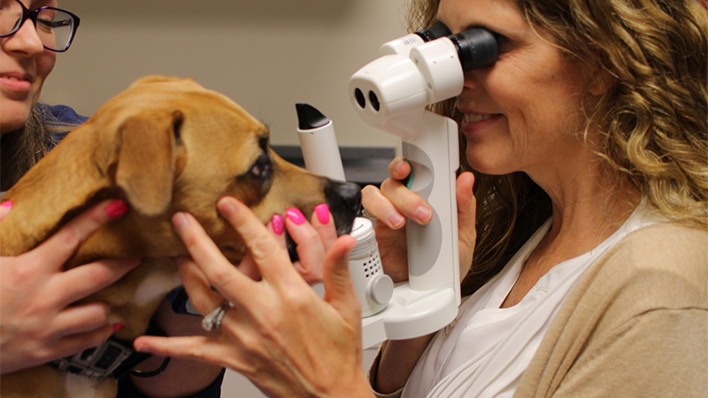
Your veterinarian will perform an eye examination to determine the location and depth of the ulcer and look for conditions that could interfere with healing. A fluorescein stain, a harmless dye that clings to damaged tissue, identifies a corneal ulcer. During a more comprehensive ophthalmic examination, your veterinarian may also evaluate tear production, eye pressure, the eyelids, and other eye structures.
If the ulcer is superficial but has failed to heal as expected, an indolent ulcer may be suspected. A veterinary ophthalmologist can provide additional evaluation and treatment when needed.
Does your dog have a nonhealing eye ulcer?
A corneal ulcer that does not heal as expected may require evaluation by a veterinary ophthalmologist. These specialists can determine why the ulcer is not healing and recommend additional treatment options to help protect your dog's eye.
Treatment for Indolent Corneal Ulcers
Because the epithelium won’t anchor to the underlying tissue, medication alone usually is not enough to make an indolent ulcer heal. Treatment focuses on removing loose or unhealthy tissue and creating a healthier surface that allows new epithelial cells to attach.
Removing Loose Corneal Tissue and Helping the Cornea Heal
The first step is often debridement—removing the loose, unattached epithelium around the ulcer so the surface can heal properly.
Debridement may be performed with a sterile cotton-tipped applicator or another technique selected by the veterinarian. For some dogs, it is combined with a procedure that stimulates healing of the corneal surface.
Diamond burr debridement is one treatment option for indolent corneal ulcers. This treatment uses a small instrument with a fine diamond-coated tip to gently remove abnormal tissue and create microscopic changes that encourage new epithelial cells to anchor.
A superficial grid keratotomy is another technique that may be used to support healing. It creates a controlled pattern of very shallow marks on the corneal surface. Despite “keratotomy,” the marks are shallow and don’t cut into the cornea.
The best approach depends on the ulcer’s characteristics and whether another eye condition is affecting healing. Your veterinary ophthalmologist will use the exam findings to recommend the safest next step.
Medications and Eye Protection
Your veterinarian may prescribe topical medications to help prevent infection and manage discomfort while the cornea heals. Pain medication may also be recommended.
A soft bandage contact lens may be placed over the eye following treatment. The lens helps protect the cornea, may improve comfort, and allows the eye to heal while still permitting prescribed medications to reach the surface.
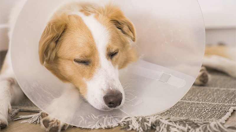
An e-collar may also be recommended to prevent your dog from rubbing or scratching the eye. Rubbing can dislodge new epithelial cells and delay healing, so protecting the eye during recovery is essential.
If your dog’s contact lens comes out, contact your veterinary team for instructions rather than attempting to replace it yourself.
"Indolent corneal ulcers can be painful and slow to heal, but appropriate treatment can help the cornea heal and protect your dog's comfort and vision."
– Vanessa J. Kuonen Cavens, DVM, DACVO
Recovery and Follow-Up
With appropriate treatment and follow-up, many dogs recover well from indolent corneal ulcers, although healing time varies. Some dogs resolve after one treatment, while others may need additional care if the ulcer remains open.
Follow-up examinations confirm the ulcer is healing as expected. Your veterinarian or veterinary ophthalmologist will monitor the cornea and determine when it has healed.
If an ulcer does not respond as expected, the eye may need to be reassessed for an underlying condition or complication that could be interfering with healing. In some cases, a more involved procedure, such as a superficial keratectomy, may be considered.
Dogs that have an indolent corneal ulcer may develop another ulcer in the same eye or the opposite eye in the future. There is no guaranteed way to prevent recurrence. If your dog develops signs of eye discomfort again, have the eye evaluated promptly so treatment can begin if another ulcer is present.
When to Seek Veterinary Care
If your dog is squinting, rubbing at an eye, tearing excessively, or has redness or a change in eye appearance, contact your veterinarian promptly. Corneal ulcers can vary in severity, and an ulcer that appears superficial can sometimes become deeper or infected.
For a persistent or nonhealing ulcer, your family veterinarian may refer you to a veterinary ophthalmologist for advanced evaluation and treatment.
Learn more ways to keep your pet healthy and safe.
About MedVet
MedVet is a nationwide network of emergency and specialty hospitals founded, owned, and led by veterinarians. Our teams provide 24/7 emergency care alongside a wide range of specialty services, with board-certified specialists collaborating across disciplines to deliver advanced, compassionate care for pets and the people who love them.
Find a MedVet location near you.
FAQs
What is an indolent corneal ulcer in dogs?
An indolent corneal ulcer is a superficial ulcer in which the outer layer of the cornea does not properly attach to the tissue beneath it. This prevents the ulcer from healing normally and can cause ongoing eye discomfort.
How are indolent corneal ulcers treated in dogs?
Treatment typically involves removing loose, unattached corneal tissue and stimulating the surface to heal. Depending on the ulcer and the dog's eye health, treatment may include debridement, diamond burr debridement, or superficial grid keratotomy, along with medications and eye protection.
How long does it take for an indolent corneal ulcer to heal in a dog?
Healing time varies between dogs. Many heal with appropriate treatment, but some dogs require additional care if the ulcer does not close as expected. Follow-up examinations help determine whether the ulcer is healing properly.
Learn More
Regular eye check-ups for your pet help maintain their eye health and detect any potential health issues.
Veterinary OphthalmologyShare




Contents
Learn More
Regular eye check-ups for your pet help maintain their eye health and detect any potential health issues.
Veterinary Ophthalmology