Glaucoma in Your Pet: Understanding and Managing This Sight-Threatening Condition
Glaucoma is a serious eye condition caused by a buildup of fluid in the eye that doesn’t drain properly, leading to increased pressure. If left untreated, it can cause blindness.
Glaucoma is a serious eye condition caused by a buildup of fluid in the eye that doesn’t drain properly, leading to increased pressure. This pressure can become painful and cause damage to the retina and optic nerve. Let’s explore the signs, types, diagnosis, and treatment options for glaucoma in pets.
What is Glaucoma in Pets?
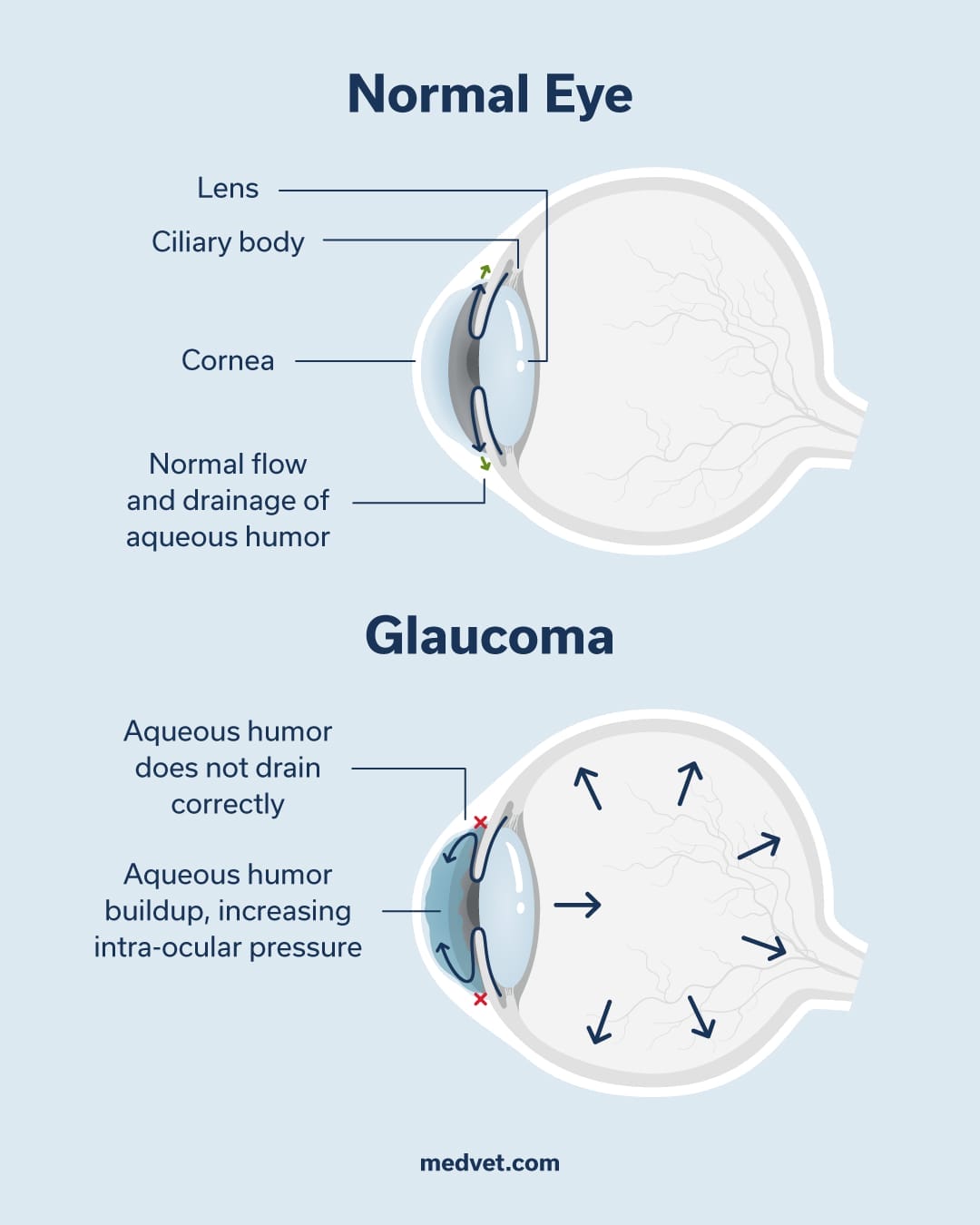
The eye contains a fluid called aqueous humor, produced by the ciliary body. This fluid fills the eye’s chambers, maintaining its shape and supplying nutrients to areas without blood supply. Glaucoma occurs when this fluid doesn’t drain correctly, causing increased pressure.
Think of it like a sink: when the drain is clear, water flows out. But if it gets clogged, water backs up. Similarly, in glaucoma, the eye’s drainage system fails, leading to fluid buildup.
Types of Glaucoma in Pets
Glaucoma in pets can be categorized into two main types: primary and secondary. Understanding the differences between these types is crucial for effective management and treatment.
Primary glaucoma is an inherited condition characterized by an abnormal drainage system in the eye. Certain dog breeds, such as Samoyed, Husky, Basset Hound, Cocker Spaniel, Beagle, Chow Chow, Maltese, Shar Pei, and Shiba Inu, are more prone to primary glaucoma due to their genetic predisposition. In cats, breeds with predisposition to glaucoma include Siamese, Burmese, and Persian cats. If your pet is diagnosed with primary glaucoma in one eye, it’s essential to monitor the other eye closely, as there’s a 50% chance it will develop glaucoma within 12 to 18 months.
Secondary glaucoma is caused by other factors, such as disease or eye injury, including conditions like uveitis, lens luxation, cataracts, retinal detachment, tumors, or trauma. This type of glaucoma can occur in any breed and is not necessarily linked to genetic factors. Identifying the underlying cause of secondary glaucoma is critical to determining the best course of treatment.
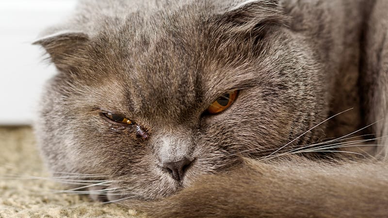
Signs of Glaucoma in Pets
Signs vary from animal to animal but may include:
- Redness around the eye
- Tearing
- Squinting
- Decreased appetite or activity
- Cloudy or bulging eye
Persistent high pressure can quickly lead to blindness—sometimes within hours in acute cases.
Diagnosing Glaucoma in Pets
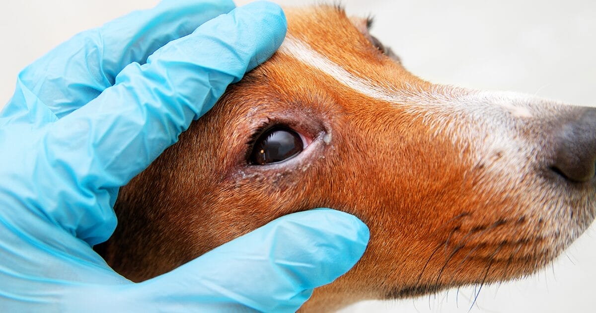
There’s no cure for glaucoma, but early diagnosis and treatment can manage the condition. A veterinary ophthalmologist will perform a detailed eye examination, including measuring intraocular pressure (IOP) using a tonometer.
Treatment aims to reduce eye pressure, typically starting with eye drop medication. Some medications decrease fluid production, while others improve drainage.
Advancements in Glaucoma Treatment for Pets
Treating glaucoma in pets has evolved significantly, offering new hope for managing this condition. Here’s an overview of the latest developments.
New Medical Therapies
Several human-approved medications are being explored for their potential in veterinary care. These include:
- Latanoprostene bunod (Vyzulta™), which improves fluid outflow and has shown promising results in lowering eye pressure in canine models.
- Netarsudil (Rhopressa™), a medication that targets the trabecular meshwork to reduce eye pressure.
- Fixed-dose combinations and sustained-release implants are also being investigated for their potential to improve treatment outcomes.
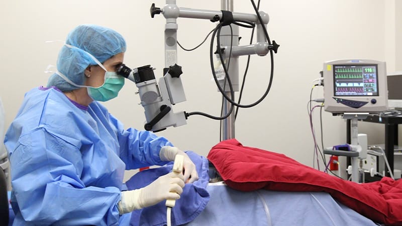
Surgical Advances
Modern surgical techniques have shown significant promise in managing glaucoma in pets. Options include:
- Ahmed valve and Baerveldt device implants, which help to drain excess fluid from the eye.
- Transscleral cyclophotocoagulation (TSCP) and Endocyclophotocoagulation (ECP), which have demonstrated high success rates for controlling eye pressure and preserving vision in dogs.
- ECP has been particularly effective, with some studies indicating that repeat procedures can extend the time before blindness occurs.
Research is ongoing into innovative approaches such as gene therapy, stem cell treatments, and neuroprotection. These emerging strategies aim to address the underlying causes of glaucoma and potentially restore or preserve vision. It’s essential to work closely with a veterinary ophthalmologist to determine the best treatment plan for your pet.
One of the challenges in managing glaucoma is ensuring pets receive their medication as prescribed. To address this, researchers are exploring sustained-release drug delivery systems that could improve treatment consistency and reduce the burden on pet owners.
Managing the Risk of Blindness and Pain from Glaucoma
If glaucoma progresses to blindness, focusing on your pet’s quality of life and managing their pain becomes essential. You can help your pet adapt to vision loss by maintaining a consistent environment, using sound cues, and enhancing scent marking. For more detailed guidance on improving a blind pet’s quality of life, you can refer to our related blog post.
Several surgical options are available to manage pain in pets with glaucoma-induced blindness. These include:
- Enucleation: Removing the eye and sewing the eyelids shut.
- Prosthetic Replacement: Removing parts of the eye and replacing them with a prosthesis for a cosmetic solution.
- Ciliary Body Ablation or Chemical Ablation: Injecting drugs to stop intraocular fluid production. This is typically reserved for blind eyes experiencing pain due to elevated pressure.
It’s essential to discuss the risks associated with each surgical option with your veterinary ophthalmologist to determine the best course of action for your pet.
Early detection and specialized care from a veterinary ophthalmologist can make a significant difference in preserving your pet’s vision. If you suspect your pet is at risk or is showing signs of eye trouble, schedule an exam with your veterinarian or a veterinary ophthalmologist promptly.
Learn more about veterinary ophthalmology.
FAQs
What is glaucoma in pets?
The eye contains a fluid called aqueous humor, produced by the ciliary body. This fluid fills the eye's chambers, maintaining its shape and supplying nutrients to areas without blood supply. Glaucoma occurs when this fluid doesn't drain correctly, causing increased pressure.
What are the signs of glaucoma in pets?
Signs vary from animal to animal but may include redness around the eye, tearing, squinting, decreased appetite or activity, and cloudy or bulging eye.
How is glaucoma in pets treated?
Treating glaucoma in pets has evolved significantly, offering new hope for managing this condition. There are multiple medical therapies available as well as surgical advances. Research is ongoing into innovative approaches such as gene therapy, stem cell treatments, and neuroprotection.
Share

Contents