Epitheliotrophic Cutaneous Lymphoma
Veterinary dermatologists investigate the causes of pets' itching and skin lesions like detectives, exploring signs and treatments.
History
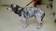
11-year-old MC Beagle presented for moderate pruritus and new skin lesions on his trunk of 2 months duration.
A hair coat color change (greying) was noted over the preceding 12 months. Cephalosporin antibiotics, antimicrobial shampoos, antihistamines, and prednisone had been dispensed without a change in lesion appearance or pruritus. Apoquel (oclacitinib) was trialed and did not reduce pruritus. Lesions continued to evolve from red skin to scale and crust to plaques and nodules. Weight loss was noted despite good appetite and interest in eating. Prior to referral, a complete blood count and serum biochemistry, urinalysis and thyroid panel did not reveal any abnormalities.

Dermatologic Examination
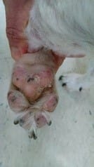
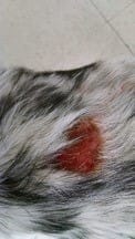
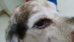
Depigmentation of the lips, eyelids, nasal planum, pawpads, and all mucocutaneous junctions. Widespread leukotrichia. Generalized exfoliative erythroderma with scale forming crusts. Multifocal variably sized alopecic, erythematous sometimes erosive or ulcerated nodules with peripheral crust on the trunk. Palpable peripheral lymphadenopathy.
Problems
Leukotrichia, nodular dermatitis, erythroderma, scale and crust, depigmentation, pruritus
Based upon the history, signalment, and lesions identified on dermatologic examination, neoplasia is the primary category of differential diagnoses to pursue. Several diagnostic tests could be recommended next. In this case, a fine needle aspiration of nodule and lymph node was done in the office as it is a rapid test with immediate results. From the nodule aspirate, there were no microorganisms, but there was a uniform population of round cells (lymphocytes). Similarly, from the lymph node there was a uniform population of round cells (lymphocytes). Based upon the results of in-office fine needle aspiration skin biopsy for dermatohistopathology was recommended to confirm the differential of epitheliotrophic lymphoma (a.k. a. cutaneous lymphoma, cutaneous T cell lymphoma, mycosis fungoides).
Histopathology of erythematous scaling skin and nodules revealed epitheliotropism, Pautrier microabscesses- pleomorphic atypical lymphocytes within the epidermis and lichenoid bands of pleomorphic lymphoid cells in the superficial dermis.
Diagnosis
Epitheliotrophic cutaneous lymphoma.
Comments
Upon examination, this case exemplifies the spectrum of lesions characteristic of cutaneous lymphoma- erythroderma, patch, plaque and tumor stages with evidence of disseminated disease (lymphadenopathy). The change in coat color over a long period of time and depigmentation at mucocutaneous junctions were key clinical features to recognize. The exfoliative erythroderma and pruritus misled the initial clinician into thinking this was a bacterial infection secondary to allergic disease. The history, signalment and distribution of lesions in this case are not supportive of allergic disease. Severe pruritus unresponsive to antipruritics is typical of cutaneous lymphoma. Although there are several diagnostic tests that would have been appropriate to pursue here given the varied lesions, I felt the most direct route to the diagnosis for this pet was FNA cytology and skin biopsy.
Pet Care Resources
For ways to ensure your pet lives a happier, healthier life, visit our Pet Care Resources library.
View ResourcesShare

Pet Care Resources
For ways to ensure your pet lives a happier, healthier life, visit our Pet Care Resources library.
View Resources